

Materials and Methods
RNA Isolation and HTS of cDNA Libraries
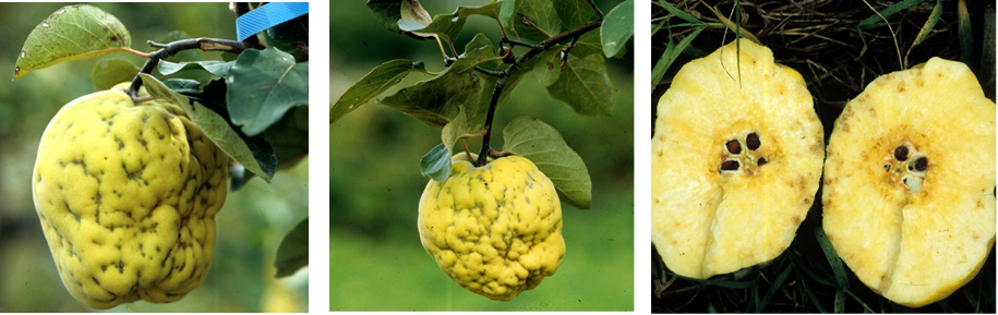
Leaf tissue, collected on 2016 from a non-symptomatic sweet orange tree (cv. Tarocco grafted on sour orange) grown in a commercial orchard in Southern Italy, was used for generating a cDNA library of small RNAs (16–30 nt). The library was generated from total nucleic acids (TNA) extracted with phenol-chloroform from leaves and sequenced (run 1 × 50) on an Illumina Genome HiScan Analyzer by Fasteris custom service (Fasteris, Switzerland) as reported previously (Di Serio et al., 2010).
Assembling of Reads and Sequencing of the Full-Length Viral Genome
Raw reads generated by HTS were filtered for quality, trimmed and de novo assembled (k-mer 15–17) using the Velvet Software 1.2.08 (Zerbino and Birney, 2008). The resulting contigs were screened by BlastX on the NCBI databases for the homologous viral sequences and those sharing significant sequence identity with CCGaV were aligned along the two RNA components of this virus, thus generating a preliminary genome scaffold of a new virus. Such a scaffold was used to design specific primers to amplify by RT-PCR overlapping cDNAs covering the full viral genome sequence (Supplementary Table S1). Amplicons were gel-purified, cloned and sequenced by Sanger Sequencing Custom Service (Macrogen, Netherlands) according to standards protocols (Navarro et al., 2018). The 5′ and 3′ termini of both CiVA genomic RNAs were determined by 5′ and 3′ rapid amplification of cDNA ends (RACE) using the specific primers reported in the Supplementary Table S1.
Sequence Analyses
RNA secondary structures were predicted by the Mfold Web Server ORF Finder at NCBI and PFAM database were used to predict the potential ORFs and identify the conserved protein domains, respectively. Modeling, prediction and analysis of CiVA proteins were performed with Phyre2 web portal When indicated, PROMALS3D was applied to generated multiple alignments of protein sequences and/or structures.
Phylogenetic Analyses
Phylogenetic trees of genomic regions, including the core amino acid sequence of RNA-dependent RNA polymerase (RdRp) and the complete amino acid sequences of movement and nucleocapsid proteins (MP and NP, respectively), were built using MEGA7 Multiple alignments were generated by Cobalt4 or Clustal Omega , then TrimAl was used to remove poorly aligned regions, thus generating a final alignment that was used to infer the phylogenetic trees adopting the maximum-likelihood method (ML) (500 bootstrap replicates). The best-fit amino acid substitution models (LG + G for RdRp and NP, and WAG + G + F for MP phylogenetic trees) were determined using MEGA7.
Bioassays, Detection, and Field Survey
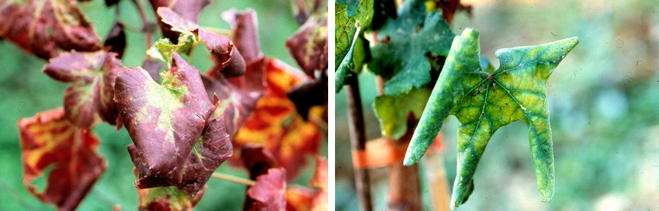
Bioassays were performed by grafting bark tissues from the CiVA-infected tree to several indicator plants, including sweet orange [Citrus sinensis (L.) Osbeck, cv. Madame vinous], grapefruit (C. paradisi Macf.), rough lemon (C. jambhiri Lush), and Dweet tangor (C. reticulata Blanco x C. sinensis). At least three plants of each species were graft-inoculated. Mock-inoculations were performed using bark tissues harvested from a certified virus-free Tarocco plant, maintained in screen-house at University of Bari.
A conventional RT-PCR assay was specifically set up for the molecular detection of CiVA. The newly developed protocol was used to carry out, during 2016 and 2017, field surveys in several orchards located in Campania and Apulia region (Southern Italy) to assess the presence and incidence of CiVA infections. The primer pair Ka-1 (5′-TCCTGATGAAGTCTTAAGATCGC-3′) and Ka-3 (5′-TTGCAGTAGTGAGAAGGGAGT-3′) was designed to amplify a cDNA fragment of 620 nt of CiVA RNA2 (Supplementary Table S1). TNA (100 ng) were extracted and reverse-transcribed as reported previously. An aliquot (2 μl) of the cDNA reaction was used for PCR amplification performed in a reaction volume of 25 μl containing 1.25 units of GoTaq polymerase (Promega, Madison, WI, United States) and a final concentration of 0.2 μM for each primer. After an initial denaturation at 94°C for 3 min, followed by 32 cycles at 94°C for 30 s, 55°C for 30 s, 72°C for 30 s and a final extension step at 72°C for 7 min, the reaction products were separated by electrophoresis on 1.4% agarose gels and visualized by UV light after ethidium bromide staining. In addition, all samples were also tested by RT-PCR for the presence of CCGaV, following the protocol described by Navarro et al. (2018).
Results
Identification of a Novel nsRNA Virus by Next Generation Sequencing
Assembly of the reads obtained through HTS of a cDNA library of small RNAs purified from leaves of a citrus tree, grown in southern Italy (Campania Region) and not showing any evident symptoms, generated a total of 7801 de novo contigs (K-mer 15 and 17). BLAST searches identified several contigs to be sequences of citrus exocortis viroid (CEVd), hop stunt viroid (HSVd), and citrus dwarfing viroid (CDVd). In addition, 35 contigs encoding deduced peptides with high amino acids (aa) sequence identity (38 to 100%) with the RdRp, the NP and the putative MP of CCGaV reported recently from citrus (Navarro et al., 2018), were also identified (Supplementary Table S2). These data suggested that a new virus, related to CCGaV, was present in the tested plant. The full-length genome of this virus, named citrus virus A (CiVA), was determined by Sanger sequencing of overlapping cDNA fragments and by 5′ and 3′ RACE.
Genomic Organization of CiVA
The CiVA genome is composed of two RNAs (RNA1 and RNA2) of 6691 and 2740 nucleotides (nt), respectively (GenBank accession numbers: MG764565 and MG764566). CiVA RNA1 and RNA2 share almost identical nucleotide sequences (up to 21 nt) at their 5′ and 3′ ends (Figure 1A). As expected for a nsRNA virus, the 5′ and 3′ termini of each genomic RNA are complementary (Bouloy, 2011) to each other, allowing the formation of a panhandle structure (Figure 1B). Interestingly, the 5′ and 3′ termini of both RNAs are identical to those of the CCGaV genomic RNAs (up to 18-nt at each terminus). Moreover, the five nt at both termini of CiVA and CCGaV genomic RNAs are identical to those of members of the family Phenuiviridae(genera Phlebovirus, Phasivirus, Tenuivirus, and Goukovirus) infecting animals and/or plants and to those of other bunyavirales-related viruses, such as the recently reported Laurel Lake virus (LLV) (Tokarz et al., 2018) infecting ticks (Figure 1C). These terminal sequences are also conserved, at least partially, in the 5′ end of the RNA1 of watermelon crinkle leaf-associated virus 1 (WCLaV-1) and watermelon crinkle leaf-associated virus 2 (WCLaV-2), which are two novel plant-infecting bunyavirales-related viruses reported in China (Xin et al., 2017; Figure 1C).
................................................................
VirFree brings together participants from both academia and privatecompanies to collaborate through their expertise on the following objectives
rfree, NGS, Nanobodies, Fruit tree viruses, viroids, virus elimination ,http://www.virfree.eu ,virfree,virfree.eu , This email address is being protected from spambots. You need JavaScript enabled to view it.


